Have you ever wondered how our bodies move and stay supported? The answer lies in the amazing human skeletal system! This complex network of bones is more than just a rigid structure; it plays a vital role in our health and is a common topic for general science sections in competitive exams like SSC, Banking, and more.
Introduction to the Human Skeletal System
What is the Human Skeletal system?
- The human skeletal system is the body’s internal framework, composed of around 270 bones at birth and 206 in adulthood.
- These bones and cartilage work together to support, move, and protect our internal organs.
- The skeleton can be divided into two main parts: the axial skeleton, which includes the skull, spine, and ribs, and the appendicular skeleton, which includes the bones of the limbs and shoulders.
- It also plays a vital role in storing minerals, like calcium, and producing blood cells.
Composition of Skeletal System
- The human skeletal system primarily comprises two main components: bones and cartilage.
- Bones are the hard, rigid structures that provide support and structure.
- Their hardness comes from a calcium-rich matrix due to calcium salts.
- Conversely, cartilage is a more flexible connective tissue found throughout the body, like the ends of bones at joints.
- There are different types of cartilage, each with its properties.
- Additionally, ligaments and tendons, which are fibrous tissues, play a crucial role in connecting bones and muscles to the skeletal system.
Composition of Human Bones
Our bones, the body’s sturdy platform, are a fascinating composite of organic and inorganic materials.
- The Collagen Network (30%): Imagine a flexible mesh – collagen, the major organic component (around 90%)! This protein, called ossein, provides tensile strength and elasticity, making bones bend without breaking.
- The Mineral Powerhouse (70%): The inorganic star is hydroxyapatite, a calcium phosphate compound. This mineral fortification gives bones their rigidity and hardness, allowing them to support our weight and protect vital organs.
- The Honeycomb Design: The internal arrangement of bone is truly ingenious. Compact bone, dense and strong, forms the outer layer. Inside, the cancellous bone resembles a honeycomb, offering lightweight support and shock absorption.
- Calcium Champion (99%) Did you know? A whopping 99% of your body’s calcium resides within bones and teeth! This mineral is crucial for bone formation and maintenance.
Important Fact about Human Bones
- The longest bone in the Human Body is the – femur (in the thigh)
- The smallest bone in the human body – Stapes (in the middle ear)
- The adult human skeleton is made up of – 206 bones
- The human infant is made up of – 270 bones
- Two principal components of bone – Collagen and Calcium phosphate
- The study of bones is called – Osteology
Understanding the 5 Types of Human Bones
The human skeleton is an architectural marvel, and the diverse shapes of its building blocks – bones – play a crucial role in its function. Let’s explore the five main bone types you’ll encounter in competitive exams:
🔹The human body consists of five types of bones. They are:
- Long Bones
- Short bones
- Flat bones
- Sesamoid bones
- Irregular bones
Long Bones
- Long bones are characterized by a central shaft, the diaphysis, flanked by two expanded ends called epiphysis.
- Comprising a dense outer layer of compact bone encasing a marrow-filled inner medullary cavity, long bones exhibit a structural balance of strength and flexibility.
- Primarily tasked with bearing body weight and facilitating movement, long bones are crucial components of the skeletal system’s framework.
- Examples of long bones include the femur, the largest and longest bone in the human body, and the ulna, a key component of the forearm.
- Notably, the femur holds the distinction of being the longest bone in the human body, contributing significantly to locomotion and structural support.
Short Bones
- Short bones exhibit dimensions where their width matches their length, resulting in a roughly cubic or cuboid shape.
- These bones play a crucial role in offering support and stability to various parts of the body.
- Examples of short bones include the carpals, forming the wrist, and the tarsals, composing the ankle.
- Due to their compact structure and role in enhancing joint flexibility and weight distribution, short bones are integral to the skeletal system’s functionality.
Flat Bones
- Flat bones exhibit a characteristic thin and curved morphology, distinguished by their flattened shape.
- Comprising three layers, flat bones feature two outer layers composed of compact bone, providing strength and protection, while an inner layer consists of spongy bone, offering flexibility and shock absorption.
- Primarily serving as protective shields for vital organs and providing ample surface area for muscular attachment, flat bones play a crucial role in safeguarding and supporting bodily structures.
- Examples of flat bones include the cranium (skull), which shields the brain, the ilium of the pelvis, the sternum, and the ribs, all contributing to the protection of vital organs and the stability of the thoracic and pelvic regions.
Sesamoid bones
- Sesamoid bones are unique skeletal elements nestled within tendons or muscles, rather than being connected to other bones by joints.
- The term “sesamoid” originates from the Arabic word “sesamum,” referring to sesame seeds, highlighting the typically diminutive size of these bones.
- An illustrative example of a sesamoid bone is the patella, commonly known as the kneecap, situated within the quadriceps tendon at the front of the knee joint.
- Sesamoid bones fulfill various functions, such as enhancing tendon mechanics, reducing friction, and providing leverage for muscle action, contributing to joint stability and efficient movement.
Irregular bones
- As their name suggests, irregular bones exhibit a shape that lacks any consistent or predictable form.
- Irregular bones frequently serve the crucial function of safeguarding delicate organs or tissues within the body.
- Examples of irregular bones include the hip bone, which forms a vital part of the pelvis, providing support and protection for the lower abdominal organs, and the base of the skull, which shields the brainstem and other essential neurological structures.
- Despite their lack of uniformity, irregular bones play indispensable roles in maintaining structural integrity and safeguarding vital bodily components.
Types of Skeletal Systems
The animal kingdom boasts a remarkable variety of body structures, and the skeletal system plays a vital role in providing support, protection, and movement. Let’s explore the two major types of skeletal systems:
Exoskeleton: The Armored Advantage (e.g., Insects, Crabs)
- Imagine wearing your armor on the outside! Exoskeletons are rigid external shells composed of chitin or calcium carbonate.
- These external frameworks offer excellent support and protection, especially for smaller animals facing predators.
- Examples include the tough exoskeletons of insects and crabs and the hard shells of snails and clams.
Endoskeleton: The Internal Architect (e.g., Humans, Vertebrates)
- In contrast to the external armor, endoskeletons are internal frameworks of cartilage and bone.
- This internal support system provides flexibility, facilitates movement, and protects vital organs.
- Vertebrates like humans, fish, birds, and amphibians showcase the strength and versatility of endoskeletons.
Difference between Endoskeleton and Exoskeleton
| Endoskeleton | Exoskeleton |
|---|---|
| Present inside the body | The present outer surface of the body |
| Animals with endoskeletons do not undergo molting. | The exoskeleton is made up of scales, chitinous cuticles, and calcified shells. |
| It is found in vertebrates. | It is mainly found in arthropods (Spiders, millipedes, centipedes, crabs, insects, etc) |
| It is a living structure. | It is a non-living structure. |
| It develops from the endoderm. | It develops from the ectoderm |
| Animals with endoskeleton do not undergo molting. | Animals with an exoskeleton are molted periodically during growth. |
Functions of Skeleton
Our skeleton, the body’s sturdy framework, plays a crucial role in keeping us healthy and functioning. Let’s explore its key functions:
Support and Protection
- The skeletal system protects the internal organs from injury by covering or surrounding them.
- The bones and cartilage form a protective cage around vital organs.
- The rib cage shields the lungs and heart, the skull (cranial bones) safeguards the brain, and the vertebral column (spine) protects the spinal cord.
Helps in Movement
- Bones act as levers, providing a firm surface for muscle attachment. Muscle contractions pull on these levers, enabling a wide range of movements.
Mineral Reservoir
- Bones are a bank for essential minerals, primarily calcium, and phosphorus.
- These minerals are stored in the bone tissue and released back into the bloodstream as needed.
- Calcium is vital for muscle function and nerve impulse transmission.
Blood Cell Production (Haematopoiesis)
- Red bone marrow, found within some bones, is the factory for red blood cells, white blood cells, and platelets.
- This process, called hematopoiesis (haemato- = “blood”, -poiesis = “to make”), ensures a continuous supply of these essential blood components.
Shape and Size
- The skeleton determines our overall body shape and size.
Balance
- The skeleton provides attachment points for muscles that help maintain balance and posture.
Hearing
Tiny bones in the middle ear (malleus, incus, stapes) transmit sound vibrations, enabling us to hear.
By understanding these functions, you gain a deeper appreciation for the amazing work your skeleton does every day!
✅ Also Read: Human Nervous System Definition, Function, Diagnostic Tests & Key Facts
Counting 206 Human bones (Table format)
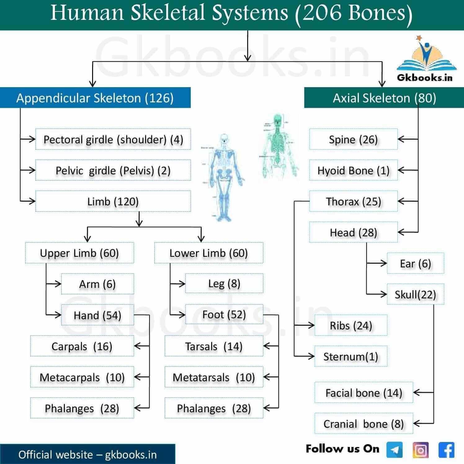
The adult human skeleton consists of 206 bones.
These 206 bones are grouped into, the axial skeleton and appendicular skeleton.
Parts of the Appendicular skeleton
- Bones of the limbs
- Shoulders
- Pelvic girdle.
Parts of Axial skeleton
- Bones of the skull
- Vertebral column
- Thoracic cage
Appendicular Skeleton
| Part of Body | Part of Endoskeleton | Region | Number of Bones | Number |
|---|---|---|---|---|
| Thorax Hip Forelimbs | Pectoral girdle Pelvic girdle | Shoulder | Scapula-Clavicle | 2 * 2 |
| Pelvis | Osinnominatum | 2 | ||
| Upper Arm | Humerus | 2 | ||
| Fore arm | Radius-Ulna | 4 | ||
| Wrist | Carpals | 16 | ||
| Palm | Metacarplas | 10 | ||
| Fingers | Phalanges | 28 * 2 | ||
| Thigh | Femur | 2 | ||
| Shank | Tibio-Fibula | 4 | ||
| Knee | Patella | 2 | ||
| Ankle | Tarsals | 14 | ||
| Sole | Metatarsals | 10 | ||
| Total | 126 | |||
Axial Skeleton
| Part of Body | Part of Endoskeleton | Region | Names of Bones | Number |
|---|---|---|---|---|
| Head | Skull | Cranium | Occipital | 1 |
| Parietal | 2 | |||
| Frontal | 1 | |||
| Temporal | 2 | |||
| Sphenoid | 1 | |||
| Ethmoid | 1 | |||
| Facial region | Nasal | 2 | ||
| Vomer | 1 | |||
| Turbinal | 2 | |||
| Lacrymal | 2 | |||
| Zygomatic | 2 | |||
| Palatine | 2 | |||
| Maxila | 2 | |||
| Mandible | 1 | |||
| Ear Ossicles | Malleus | 2 | ||
| Incus | 2 | |||
| Stapes | 2 | |||
| Back bone | Vertebral column | Hyoid | Hyoid body | 1 |
| Neck | Cervical vertebrae | 7 | ||
| Thorax | Thoracic vertebrae | 12 | ||
| Waist | Lumber vertebrae | 5 | ||
| Sacrum | Sacral vertebra Sacrum | 1 | ||
| Tail | Caudal vertebrae / Coccyx | 1 | ||
| Thorax | Sternum Ribs | — | Sternum | 1 |
| — | Ribs | 24 | ||
| Total | 80 | |||
| Total Bone = Appendicular Skeleton + Axial Skeleton = 126 + 80 = 206 |
|---|
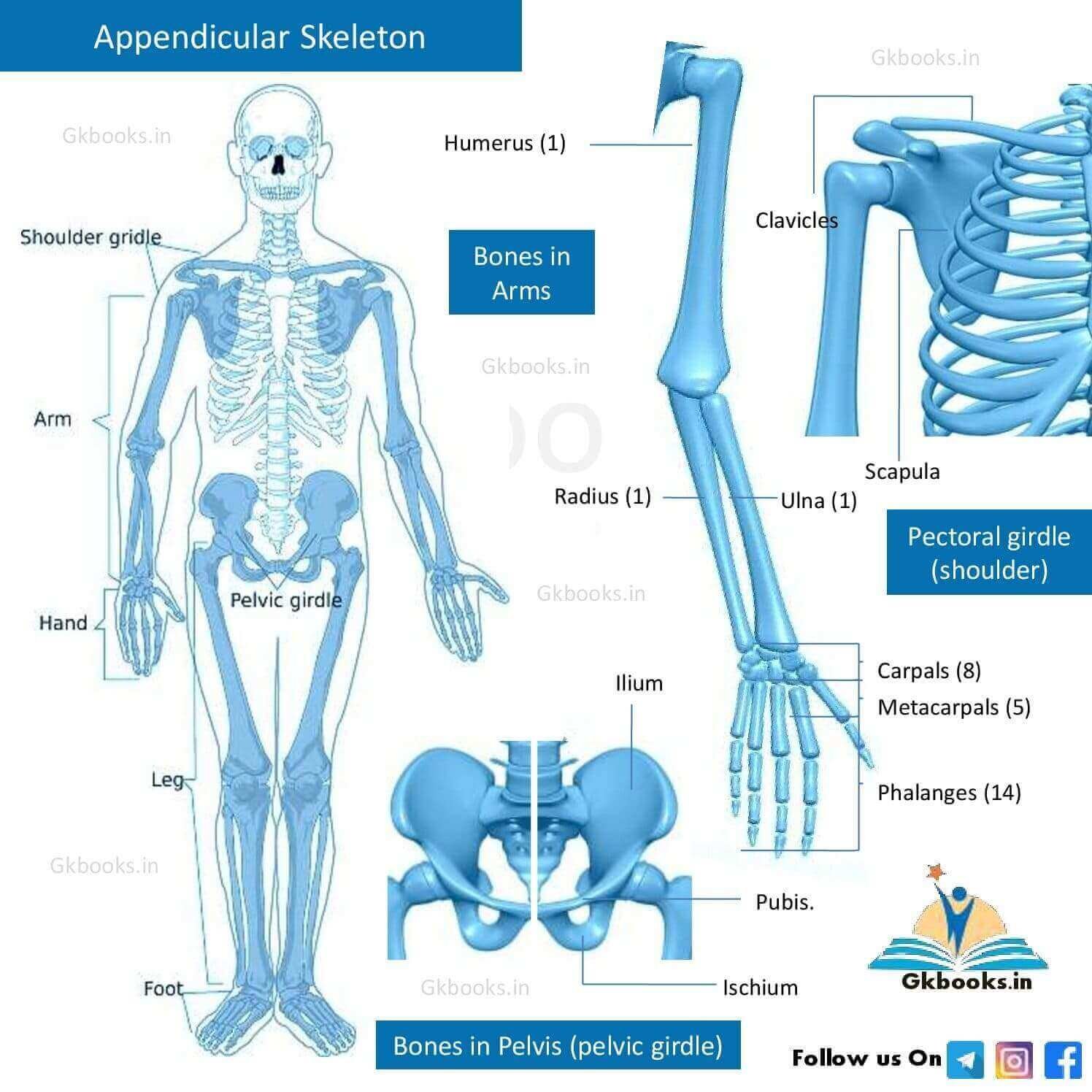
Diagram of Axial and Appendicular Skeletal System of Human
Counting 206 Human bones (Simplified format)
Appendicular Skeleton
• The appendicular skeleton, comprising the arms and legs, including the shoulder and pelvic girdles, contains 126 bones.
■ Bones in Arms
◘ Human Arms consist of 64 bones (32 in each arm)
◘ Bones in Upper Arm [ 3 × 2 = 6 bones , 3 on each side]
☞ Humerus – 2
♦ Under Pectoral girdle (shoulder)
☞ Scapula – 2
☞ Clavicles – 2
◘ Bones in Lower Arm( 2× 2 = 4 bones, 2 on each side)
☞ Ulna – 2
☞ Radius – 2
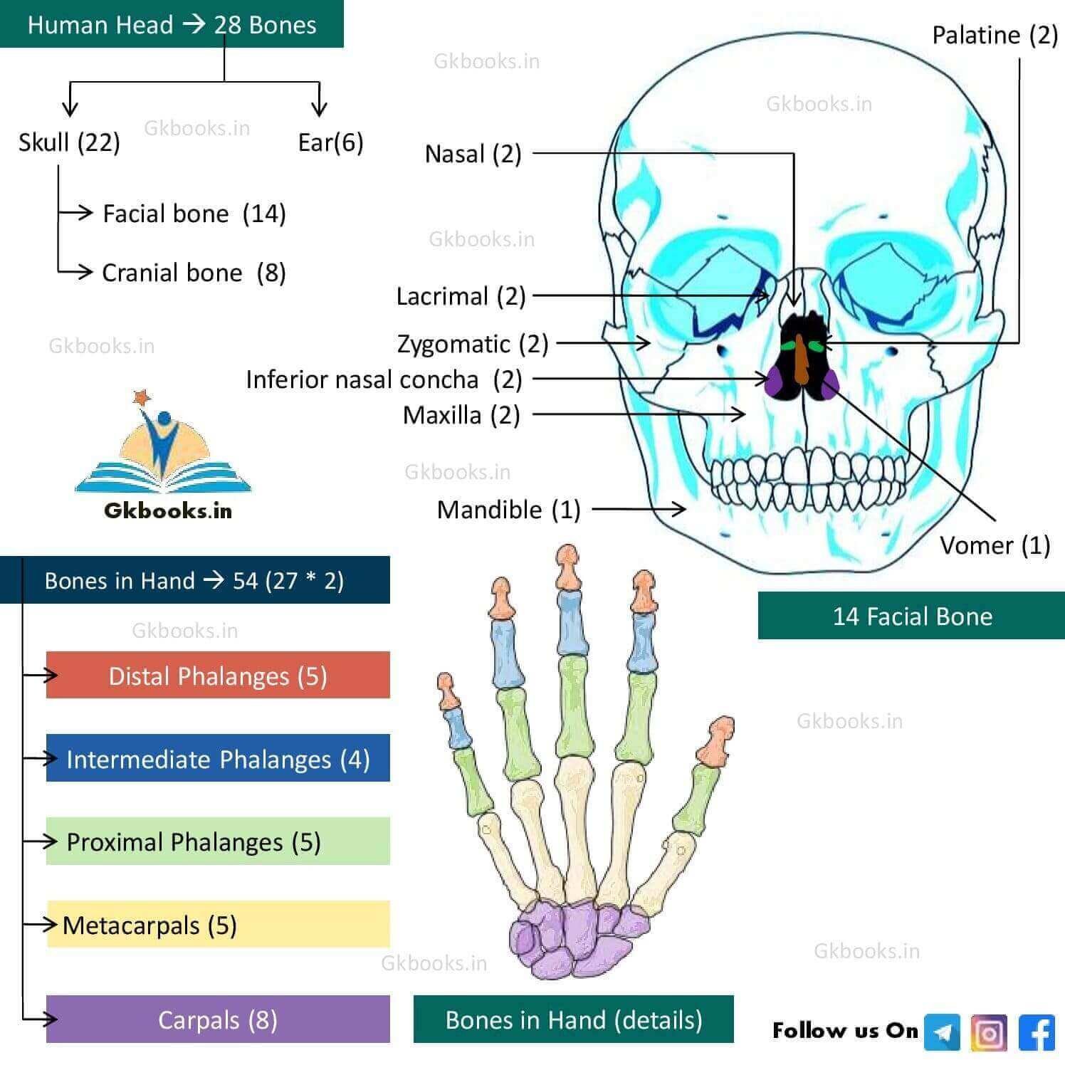
◘ Bones in Hand (27 × 2 = 54 bones, 27 in each hand)
♦ Under Carpals
☞ Scaphoid bone – 2.
☞ Lunate bone – 2.
☞ Triquetral bone – 2
☞ Pisiform bone – 2
☞ Trapezium – 2
☞ Trapezoid bone – 2
☞ Capitate bone – 2
☞ Hamate bone – 2
☞ Metacarpals ( 5 × 2 =10 bones, 5 on each side)
♦ Under Phalanges of the hand
☞ Proximal phalanges ( 5 × 2 =10 bones, 5 on each side)
☞ Intermediate phalanges (4 × 2 = 8 bones, 4 on each side)
☞ Distal phalanges (5 × 2 = 10 bones, 5 on each side)
✪ Total = 6 (upper arm) + 4 (lower arm) + 54 (both hands) = 64
■ Bones in Pelvis (pelvic girdle)
☞ The Pelvis region contains Two coxal bones.
☞ These 2 coxal bones are made up of 3 regions (fused): ilium, ischium, and pubis.
■ Bones in Leg
◘ There are a total of 30 × 2 = 60 bones , 30 in each leg.
☞ Femur – 1× 2 = 2 bones
☞ Patella or kneecap – 1× 2 = 2 bones
☞ Tibia – 1× 2 = 2 bones
☞ Fibula – 1× 2 = 2 bones.
♦ Bones in Foot (26 × 2 = 52 bones, 26 per foot)
♦ Human foot consists of Tarsals, Metatarsals and phalanges
➧ Bones in Tarsals (7 × 2 = 14 bones, 7 in each Tarsals )
☞ Calcaneus or heel bone – 1 × 2 bones
☞ Talus – 1 × 2 = 2 bones
☞ Navicular bone – 1 × 2=2 bones
☞ Medial cuneiform bone – 1 × 2=2 bones
☞ Intermediate cuneiform bone – 1 × 2 =2 bones
☞ Lateral cuneiform bone – 1 × 2=2 bones
☞ Cuboid bone – 1 × 2 = 2 bones
➧ Bones in Metatarsals – 10 bones
➧ Bones in Phalanges of the foot (14 × 2= 28 bones, 14 in each Phalanges )
☞ Proximal phalanges – 5 × 2 = 10 bones
☞ Intermediate phalanges – 4 × 2 = 8 bones
☞ Distal phalanges – 5 × 2 = 10 bones
Axial Skeleton
The axial skeleton, comprising the spine, chest, and head, contains 80 bones.
■ Bones in Spine
◘ A fully grown adult has 26 bones in the spine.☞ Cervical vertebrae – 7 bones
☞ Thoracic vertebrae – 12 bones
☞ Lumbar vertebrae – 5 bones
☞ Sacrum – 1 bones (Adult)
☞ Coccygeal vertebrae – 1 bone
■ Bones in Chest (Thorax)
◘ There are 25 bones in the chest.
☞ Sternum – 1
☞ Ribs – 24, in 12 pairs
■ Hyoid Bone – 1
■ Bones in Skull
◘ The human skull made up of 22 bones
◘ The Human Head contains 28 bones, including the middle ear bones.
| 28 bones in Head ➥ 22 + 6 = 28 | ||
|---|---|---|
| 22 bones in Skull ➥ 14 + 8 = 22 | 6 bones in Ear | |
| 8 Cranial Bones | 14 Facial Bones | 3 × 2 = 6 Bones |
| • Occipital bone • Parietal bones – 2 • Frontal bone • Temporal bones – 2 • Sphenoid bone • Ethmoid bone | • Nasal bones – 2 • Maxillae – 2 • Lacrimal bone – 2 • Zygomatic bone – 2 • Palatine bone – 2 • Inferior nasal concha – 2 • Vomer – 1 • Mandible – 1 | • Malleus – 2 • Incus – 2 • Stapes – 2 |
Human Skeletal System flow chart of 206 bones
Disorders of Human Skeletal Systems
| Disease | Description |
|---|---|
| Osteoporosis | It is an age-related disorder in which bones lose mineral density, weaken, and break more easily than normal bones. |
| Osteoarthritis | Rickets is a skeletal disorder in which the development of bone is disrupted. The lack of vitamin D. causes rickets It is a common disease in children. It causes bone pain, poor growth, and soft, weak bones that can lead to bone deformities |
| Paget’s disease | Paget’s disease is a chronic bone disorder that causes affected bones to become large and misshapen. |
| Bursitis | Bursitis is the painful swelling of a small, fluid-filled sac called a bursa due to physical injury. The most common locations for bursitis are in the shoulder, elbow, and hip. |
| Rickets | Bursitis is the painful swelling of a small, fluid-filled sac called a bursa due to physical injury. The most common locations for bursitis are in the shoulder, elbow, and hip |
| Osteomalacia | Osteomalacia is the most common nutritional deficiency among adults. Osteomalacia is a disorder of “bone softening” that occurs due to a prolonged deficiency of vitamin D. |
Source:
- Encyclopedia of General Science Book
Explore More Related Posts:
Structure, Components, and Functions of Cell: A Complete Overview
Modes of Nutrition in Living Organisms with Chart and Tables
Human Nervous System Definition, Function, Diagnostic Tests & Key Facts
Human Immune System: Immunity, Lymphocytes, Antibody with Diagrams
Steps Involved in Light Reaction of Photosynthesis With Short Question


